Eyes
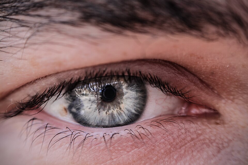

The eye is a round organ for sensing light so organisms can see. It is the first part of the visual system. About 97 percent of animal species have eyes. Image-resolving eyes are present in cnidaria, molluscs, vertebrates, annelids and arthropods.
In mammals, two kinds of cells, rods and cones, allow sight by sending signals through the optic nerve to the brain.
Some animals can see light that humans cannot see. They can see ultraviolet or infrared light.
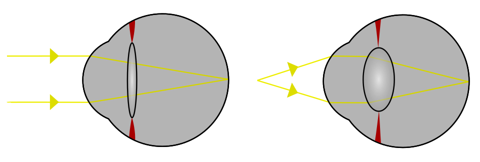
The lens on the front part of the acts like a camera lens. It can be pulled flatter by muscles inside the eye, or allowed to become rounder. As some people get older, they may not be able to do this perfectly. Many people are born with other small problems or get them later in life. Eyeglasses (or contact lenses) may fix the problem.
Like different cameras, different eyes have different abilities. They may have higher or lower resolution, the ability to detect small details. They may have different performance in low light; nocturnal animals can see better at night than daytime animals. They may have different ability to distinguish colours.
Parts of the eye
The human eye is composed of several different parts. These parts may or may not be the same in other animals. They are:
- Cornea: the outermost, transparent layer that protects the iris and pupil.
- Pupil: the black circle in the middle of the eye, through which light passes.
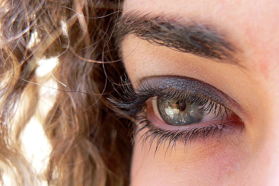
- Iris: the colorful circle of the eye around the pupil. It can be brown, blue, green, etc. Its main function is to regulate the amount of light coming into the eye.
- Sclera: the large, white field around the iris that keeps the eyeball in shape.
- Lens: behind the cornea there is a transparent bioconvex lens of very short focal length.This lens is held in the centre of eye ball with the help of ciliary muscles.
- Retina: has the cells which turn light into nerve impulses.
- Optic nerve: the nerve that connects the eye to the brain. Optical information is taken to the rear of the brain for processing: see cerebral cortex.
Types of eye
Today, ten different types of eyes are known. Most ways of capturing an image have evolved at least once.
One way to categorize eyes is to look at the number of “chambers”. Simple eyes are made of only one concave chamber, perhaps with a lens. Compound eyes have many such chambers with their lenses on a convex surface.
Eyes also can be grouped according to how the photoreceptor is made. Photoreceptors are either ciliated, or rhabdomic. and some annelids possess both.
Pit eyes
Pit eyes are set in a depression in the skin. This reduces the angles at which light can enter. It allows the organism to say where the light is coming from.
Such eyes can be found in about 85% of phyla. They probably came before the development of more complex eyes. Pit eyes are small. They are made of up to about hundred cells, covering about 100 µm. The directionality can be improved by reducing the size of the opening, and by putting a reflective layer behind the receptor cells.
Pinhole eye
The pinhole eye is an advanced form of pit eye. It has several bits, most notably a small aperture and deep pit. Sometimes, the aperture can be changed. It is only found in the Nautilus. Without a lens to focus the image, it produces a blurry image. Consequently, nautiloids can not discriminate between objects with a separation of less than 11°. Shrinking the aperture would produce a sharper image, but let in less light.
Spherical lensed eye
The resolution of pit eyes can be improved a lot by adding a material to make a lens. This will reduce the radius of the blurring, and increase the resolution that can be achieved. The most basic form can still be seen in some gastropods and annelids. These eyes have a lens of one refractive index. It is possible to get a better image with materials that have a high refractive index which decreases towards the edges. This decreases the focal length and allows a sharp image to form on the retina.
This eye creates an image that is sharp enough that motion of the eye can cause significant blurring. To minimize the effect of eye motion while the animal moves, most such eyes have stabilizing eye muscles.
The ocelli of insects have a simple lens, but their focal point always lies behind the retina.They can never form a sharp image. This limits the function of the eye. Ocelli (pit-type eyes of arthropods) blur the image across the whole retina. They are very good at responding to rapid changes in light intensity across the whole visual field — this fast response is accelerated even more by the large nerve bundles which rush the information to the brain. Focusing the image would also cause the sun’s image to be focused on a few receptors. These could possibly be damaged by the intense light; shielding the receptors would block out some light and reduce their sensitivity.
This fast response has led to suggestions that the ocelli of insects are used mainly in flight, because they can be used to detect sudden changes in which way is up (because light, especially UV light which is absorbed by vegetation, usually comes from above).
Refractive cornea
The eyes of most land-living vertebrates (as well as those of some spiders, and insect larvae) contain a fluid that has a higher refractive index than the air. The cornea is sharply curved and refracts light towards the focus. The lens need not do all of the refracting. This lets the lens adjust the focus more easily, for much higher resolution.
Reflector eyes
Instead of using a lens it is also possible to have cells inside the eye that act like mirrors. The image can then be reflected to focus at a central point. This design also means that someone looking into such an eye will see the same image as the organism which has them.
Many small organisms such as rotifers, copeopods and platyhelminthes use such this design, but their eyes are too small to produce usable images. Some larger organisms, such as scallops, also use reflector eyes. The scallop Pecten has up to 100 millimeter-scale reflector eyes fringing the edge of its shell. It detects moving objects as they pass successive lenses.
Compound eyesCompound eyes
Compound eyes are different from simple eyes. Instead of having one organ that can sense light, they put together many such organs. Some compound eyes have thousands of them. The resulting image is put together in the brain, based on the signals of the many eye units. Each such unit is called ommatidium, several are called ommatidia. The ommatidia are located on a convex surface, each of them points in a slighly different direction. Unlike simple eyes, compound eyes have a very large angle of view. They can detect fast movement, and sometimes the polarization of light.
Compound eyes are common in arthropods, annelids, and some bivalved molluscs.
Evolution of the eye
The evolution of eyes started with simplest light-sensitive patches in unicellular organisms. These eye-spots do nothing but detect if the surroundings are light or dark. Most animals have a biochemical ‘clock’ inside. These simple eye-spots are used to adjust this daily clock, which is called circadian rhythm. Some snails, for example, see no image (picture) at all, but they sense light, which helps them stay out of bright sunlight.
More complex eyes have not lost this function. A special type of cells in the eye senses light for a different purpose than seeing. These cells are called ganglion cells. They are located in the retina. They send their information about light to the brain along a different path (the retinohypothalamic tract). This information adjusts (synchronizes) the animal’s circadian rhythm to nature’s light/dark cycle of 24 hours. The system also works for some blind people who cannot see light at all.
Eyes that are a little bit better are shaped like cups, which lets the animal know where the light is coming from.
More complex eyes give the full sense of vision, including color, motion, and texture. These eyes have a round shape that makes light rays focus on the back part of the eye, called the retina.
Other
Good fliers like flies or honey bees, or prey-catching insects like praying mantis or dragonflies, have specialized zones of ommatidia organized into a fovea area which gives sharp vision. In this zone the eyes are flattened and the facets are larger. The flattening allows more ommatidia to receive light from a spot. This gives a higher resolution.
The body of Ophiocoma wendtii, a type of brittle star, is covered with ommatidia, turning its whole skin into a compound eye. The same is true of many chitons.
Sign and symptoms of disease or illness
pupil: Argyll Robertson pupils (picture)
Adie pupil (picture)
Marcus Gunn pupil
cornea: Fleischer ring
Kayser–Fleischer ring
Hudson–Stahli line
iris: Brushfield spots
Lisch nodule
conjunctiva: Bitot’s spots
Arlt’s line
retina: Hollenhorst plaque
Roth’s spot
Fuchs spot
others: Alexander’s law
Hirschberg test
Siegrist streaks
eyelid: Abadie’s sign of exophthalmic goiter
Boston’s sign
Dalrymple’s sign
Stellwag’s sign
lacrimal: Schirmer’s test
Written for younger readers
Eyes, in simpler words
This version comes from Wikijunior, a set of books written for children aged 8 to 11. It is shorter and uses plainer language than the article above.
From Wikijunior: Human Body
The eye is a very perplexing system that allows us to view things. Here you will find a lot about the eye. But remember, this is not a professional page, but rather an outline.
Eyes are the parts of our body that perceive light. They allow us to see the world and to understand how objects relate to each other. We can distinguish far objects from close ones and determine their color and shape. The eye acts like a camera where the eye-lens is a replacement of camera-lens, eye-lid of shutter and retina of photographic film.
The eye is almost spherical in shape. The eye-ball is about of 2.3cm diameter. We perceive eyes together with eyelids and eyebrows which make up one main characteristic of the face.
Eyes are made of several different parts. The white part of the eye is called the sclera.(It is interesting to note that only in Human Species among all other creatures that sclera is visible when the eyes are open). This part covers the surface of the eyes except at the very front. At the front of an eye is a cornea. This part acts like a shield for the eye. It can still let light pass through because it is transparent. Under the cornea is the iris and pupil. The pupil is the black circle in the middle. It is actually a hole that lets light pass through into the back of the eye. The iris is a coloured diaphragm around the pupil. It is a muscle that opens and closes around the pupil to let more or less light through. Behind the pupil is a lens that focuses the light that passes through it onto the retina. It is made up of jelly-like crystalline substances and is convex in nature. Between the lens and cornea is a fluid called aqueous humour.Similarly, inside the major part of eye ball is vitreous humour.
The retina is the lining of the inside back of the eye. It is the part of the eye that translates the information from the light into the language of the brain i.e. electric impulses. The retina is lined with thousands of tiny rods and cones which detect the color and amount of light shining onto them.
The optical information gathered by the rods and cones is passed via the optic nerve at the back of the eye to the brain.
Most of the reflection takes place through cornea, aqueous humour and vitreous humour and the lens merely function to focus the image on retina. The lens is held by ciliary muscles which also controls the curvature of lens to accommodate it to see distant and close objects clearly.
The brain processes the raw data from the eyes to make sense of what you see compared to your knowledge of the world around you. The brain interprets what it receives from the eyes. You do not directly “see”.
A clear example of this can be seen from experiments where subjects wear glasses that invert the view (turn it upside down). After a while the brain will adjust and turn the scene back up the right way because that agrees with other more reliable information you are receiving.
Similarly the brain adjusts changes in color and light levels to better match how the objects should appear.
It also combines the two slightly different views from your left and right eyes to work out the distance of objects from you. Your right eye will show a little more of an object’s right side and the left eye will show more of an objects’ left side. This is called stereoscopic vision. This vision helps us to judge the distance accurately and provides us a 3D vision of the world. The difference between the views from each eye becomes less the further the object is away from you.
Our perceptions are not perfect and can lead to misinterpretations of what we see. These are called optical illusions.
However even though the brain’s processing can be inaccurate, at most times we perceive a faithful reconstruction of the real world. Only optical illusions remind us that there is at times a difference between perception and the true state of affairs. Without our eyes and the complex processing of visual information by our brains we would not be able to make sense of writing, art or photos, nor understand as much as we do from limited visual information.
The optic nerve connects the eyes with the brain. Several eye muscles (4 rectus and 2 oblique muscles) attach the eyes to the skull and perform eye movements to control our gaze and visual focus and the power of accommodation of our eyes.
You should avoid putting your hands or any other object in the eyes. Wearing safety goggles when doing construction work can protect your eyes from injury. You should not watch television or use a computer for long periods of time. You should keep books at a distance of about 25cm while reading. Avoid looking at bright objects (like the sun) for too long as it can damage your eyes and even make you blind.
ja:Wikijunior:人間の体/目
Where this page comes from
The article above is adapted from “Eyes” on Simple English Wikipedia, by its contributors. We removed reference markers, navigation boxes and tables, expanded measurement templates into readable numbers, and kept the prose otherwise intact. The simpler version is adapted from Wikijunior on Wikibooks.
Both sources are published under CC BY-SA 4.0, so this page is published under the same licence. You may share and adapt it, including commercially, as long as you credit the original and keep the same licence.
Worksheets, answer keys and printable layouts elsewhere on K5Print are our own work and are not covered by this licence.