Histology
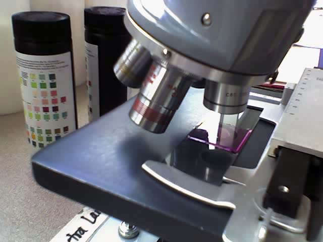

Histology is the study of the microscopic anatomy of cells and tissues of plants and animals, particularly the tissues. It is a part of cytology, and an essential tool of biology and medicine.
Histology is usually done by looking at cells and tissues under a light microscope or electron microscope. The tissue has to be specially prepared beforehand.
The process
The stages below are only described in outline. Laboratories which do histology work from schedules which are much more detailed.
Fixing
Chemical fixatives are used to preserve tissue from decay. This preserves the structure of the cell and of sub-cellular components such as cell organelles (e.g., nucleus, endoplasmic reticulum, mitochondria). The most common fixative for light microscopy is formalin (4% formaldehyde in saline).
Embedding
After fixing, the block of tissue is embedded in paraffin wax. This holds and preserves the tissue as a block.
Sectioning
The section is cut into a series of wafer-thin slices, each of which is put on a glass microscope slide. The machine which cuts the block is a mechanical guillotine which can be set to cut at a suitable depth for the tissue in question.
Staining
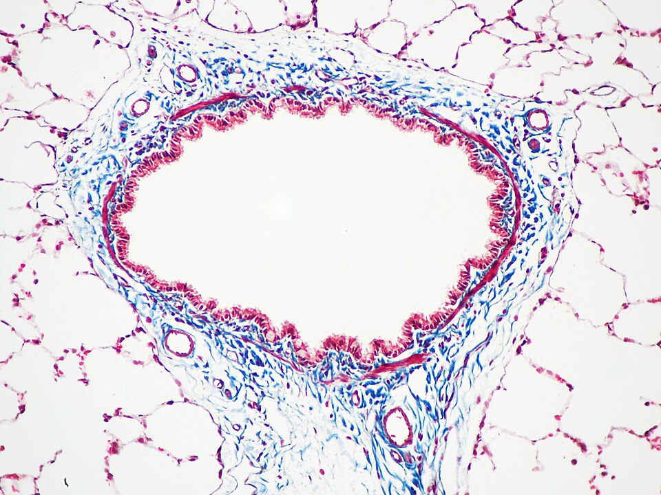
Stains are dyes, chemicals used to make cells and tissues easy to see under a microscope. There are many tissue stains, and each of them has advantages and disadvantages.
Haematoxylin and eosin (H&E)
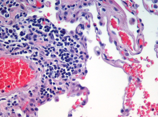
This is the most widely used stain in biology and medicine. Haematoxylin colours cell nuclei and eosin colours cell cytoplasm.
Silver nitrate
Camillo Golgi developed a silver nitrate stain for nerve cells. His idea was used by Santiago Ramón y Cajal in his famous work on the structure of brain tissue.
Modern techniques
Electron microscopy is used often as well as light microscopy. This has its own procedures. Its advantage is that it resolves things which light cannot resolve. For example, viruses were first seen by electron microscopy..
Specialised selective stains using immunology or radioactive labelling are now routine. The advantage of using antibodies or radioactive labels is that they stick to specific kinds of molecules. Increasingly popular is tagging with a fluorescent stain, which shows up even if a tiny part of a cell is stained. Immunoinfluorescence is the name of this particular technique.
Where this page comes from
The article above is adapted from “Histology” on Simple English Wikipedia, by its contributors. We removed reference markers, navigation boxes and tables, expanded measurement templates into readable numbers, and kept the prose otherwise intact.
Both sources are published under CC BY-SA 4.0, so this page is published under the same licence. You may share and adapt it, including commercially, as long as you credit the original and keep the same licence.
Worksheets, answer keys and printable layouts elsewhere on K5Print are our own work and are not covered by this licence.