Lungs
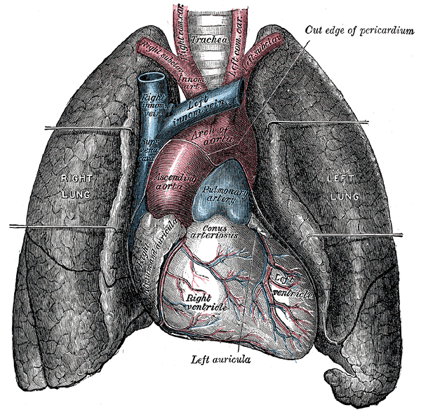

The lung is an organ in many vertebrates (animals having a spine, or backbone). It takes blood oxygen from the air, and expels carbon dioxide. Most vertebrates with lungs have two of them.
In animals, the lungs are the area where gas exchange takes place. Without gas exchange, oxygen would not pass into the blood from the lungs so the body cells would not be able to receive the oxygen needed for respiration.
Gas exchange happens in the alveoli. The alveoli are moist to allow oxygen to move from the lung through the alveoli into blood vessels and red blood cells. Carbon dioxide passes from the blood into the alveoli. The oxygen-filled blood goes back to the heart and the carbon dioxide in the alveoli is pushed out of the lungs and into the air we breathe out.
Bird lung
Birds lungs are smaller than mammal lungs, and do not have alveoli, instead they have millions of para-bronchi. These para-bronchi end up in tiny capillaries or very small blood vessels and they pass close to the body’s blood vessels, so diffusion can occur and the oxygen and carbon dioxide is exchanged. The oxygen and carbon dioxide in birds lungs are continuously diffused into and out of the blood, not like in mammals where diffusion can only happen in the alveoli. Air does not go into and back out of the lungs as in mammals. Instead, muscular air sacs push the air forward through bird lungs.
Reptile lung
Reptile lungs open and close because of the ribs surrounding them pressing down on them and then opening up with the help of muscles. The liver is also attached to the bottom of the lungs and when a muscle which is attached to the liver pulls, the liver moves away from the lungs and pulls them, making them bigger.
Amphibian lung
Frog lungs are very simple compared to most other lungs, they are simply balloons, with moist outsides allowing for diffusion. But frogs do not move around much and so do not need lots of oxygen, but they can also take in oxygen through their moist outer skin if a big demand of oxygen is needed (e.g. Fight or flight response)
Written for younger readers
Lungs, in simpler words
This version comes from Wikijunior, a set of books written for children aged 8 to 11. It is shorter and uses plainer language than the article above.
From Wikijunior: Human Body
NOTOC The lung is an organ we use when we breathe. It takes oxygen from the air and transports it to the blood. It also takes carbon dioxide from the blood and releases it into the air.
Your lungs are located in the chest on either side of the heart. There are two of them. They are similar in appearance, but not identical.
Lungs have a spongy texture and a honeycomb pattern. They have much more surface area in total than just the outer surface of the lung. Each lung is separated into lobes. There are three lobes on the right and two on the left. The left lung contains a dent called the “cardiac notch”. The cardiac notch allows room for the heart. The lungs form the major part of thoracic cavity.
The two main bronchi are the tubes that enter the roots of the lungs. The bronchi continue to divide within the lung with each branch smaller than the last. At the end of the smallest bronchi (called terminal bronchioles) there are tiny sacks. These sacks are made up of clusters of alveoli, which look a little like grapes in a bunch. The individual alveoli are wrapped in blood vessels. This is where the gas exchange actually occurs. Blood without much oxygen from the heart is pumped through the pulmonary artery to the lungs. Oxygen from the air diffuses into blood and carbon dioxide from the blood is released. The oxygen-rich blood returns to the vein via the pulmonary veins to be pumped back into the rest of the body. The process goes on until you die.
The cells in our body need oxygen to produce energy. These same cells produce carbon dioxide as a by-product. So there has to be a way for the body to deliver oxygen to cells and carry carbon dioxide from cells. The lungs allow your body to trade its waste carbon dioxide for oxygen.
The lungs also serve as a cushion or shock absorber for the heart.
The diaphragm is a big muscle under your lungs. It pushes downward when you inhale, which enlarges the cavity in the chest and causes the lungs to expand. This causes air to flow into them. When the diaphragm relaxes, air flows out and you exhale. One name for this process is ventilation.
The lungs directly connect the respiratory system to the circulatory system, for in the lungs, oxygen will leave the air and enter the blood so that it can be transferred to all of the other cells in your body. Conversely, carbon dioxide is removed from blood back into the air, so that it does not poison the cells that produced it.
Breathing is largely driven by the muscular diaphragm at the bottom of the thorax. Contraction of the diaphragm pulls the bottom of the cavity in which the lung is enclosed downward. Air enter through the oral and nasal cavities; it flows through the larynx and into the trachea, which branches out into bronchi. Relaxation of the diaphragm has the opposite effect, passively recoiling during normal breathing. During exercise, the diaphragm contracts, forcing the air out more quickly and forcefully. The rib cage itself is also able to expand and contract to some degree, through the action of other respiratory and accessory respiratory muscles. As a result, air is sucked into or expelled out of the lungs, always moving down its pressure gradient. This type of lung is known as a bellows lung as it resembles a blacksmith’s bellows. Lungs have a tremendous reserve volume as compared to the oxygen exchange requirements when at rest.
Vital capacity is the maximum volume of air that a person can exhale after maximum inhalation. A person’s vital capacity can be measured by a spirometer (spirometry). In combination with other physiological measurements, the vital capacity can help make a diagnosis of underlying lung disease.
The environment of the lung is very moist, which makes it hospitable for bacteria. Many respiratory illnesses are the result of bacterial or viral infection of the lungs.
To keep your lungs healthy, you should not smoke cigarettes and avoid smoky environments. You should not work or live in environments where the air is contaminated with dust or chemicals. If you do have to go into those kinds of environments, wear a respirator. Aerobic exercise like swimming and running can increase your lungs’ vital capacity. If the alveoli can’t get enough air, a person may have asthma.
Where this page comes from
The article above is adapted from “Lungs” on Simple English Wikipedia, by its contributors. We removed reference markers, navigation boxes and tables, expanded measurement templates into readable numbers, and kept the prose otherwise intact. The simpler version is adapted from Wikijunior on Wikibooks.
Both sources are published under CC BY-SA 4.0, so this page is published under the same licence. You may share and adapt it, including commercially, as long as you credit the original and keep the same licence.
Worksheets, answer keys and printable layouts elsewhere on K5Print are our own work and are not covered by this licence.